Why dna is double helix?

0

DNA is Double Helix

Erwin Chargaff and his colleagues at Columbia University in 1940’s observed that four bases in DNA occur in definite proportions and concluded:
  • DNA specimen isolated from different tissues of the same species has the same base composition.
  • The base composition of DNA varies from one species to another.
  • The base composition of DNA in a given species doest not change with the age of organism, its nutritional status, or change in environmental conditions.
  • The number of adenine residues in all DNA’s regardless of the species is equal to the number of thymine residues (i.e. A=T), and the number of guanine residues is equal to the number of cytosine residues (C=G). This led to Chargaff’s rule.
  • We find in the above table that the amount of A in yeast is nearly equal to T, the slight error may be an experimental one.
In 1953, nearly 20 years after James Watson and Francis Crick gave the three dimensional modal for DNA using Chargaff’s rule and X-Ray diffraction studies made by Rosalind Franklin and Mauric Wilkins. In 1962, Watson, Crick and Wikins got Nobel’s prize for elucidating the structure of DNA.

Watson and Crick -DNA Model

DNA model as proposed by Watson and Crick

  • Two right handed helical polynucleotide chains are coiled around a common axis and are running in opposite direction.
  • The purine and pyrimidine bases are on the inside of the helix, whereas phosphate and deoxyribose units are on the outside. The plane of the bses is perpendicular to the axis. The planes of the sugar are nearly at right angle to those of the bases.
  • The diameter of helix is 20Ao. Adjacent bases are separated by 3.4Ao along the helix axis and related by a rotation of 36o. The helical structure repeats after every ten residues on each chain i.e. at an interval of 34Ao.
  • Two chain are held together by hydrogen bond between pair of bases. Adenine is always paired with thymine and guanine with cytosine.
hydrogen bonding between adenine and thymine
Hydrogen bonding between adenine and thymine
hydrogen bonding between cytosine and guanine
Hydrogen bonding between cytosine and guanine
The Watson-Crick model is also referred to as B-DNA. This form of the DNA is the most stable structure for random sequences. DNA can occur in three different conformations. Two other variant DNA structures, that have been well characterized in crystal structures, are the A and Z-DNA.
Watson and Crick model of DNA

Types of DNA

A-DNA: It is a favored DNA structure in many solutions that are relatively devoid of water. It is right handed helical structure with rise in base pairs 2.3Ao and number of bases per helical turn in 11. It is shorter and thicker than B-DNA and has a central hole. Z-DNA. Unlike B and A-DNA, Z-DNA is left handed and has 12 base pairs per turn, the rise in base pairs is 3.8Ao. This type of DNA is observed when there is repeated sequence of C and G. This repeat leads to Zigzag DNA. Palindrome sequences: Sometime in DNA the base sequence on one strand repeats on the other strand or invert repeat, is called palindrome sequence as shown. They have two-fold axis of symmetry. They are the important points of attack by restriction endonucleases in DNA molecules.
palindrome sequences
Palindrome sequences
Forces Stabilizing the DNA Covalent bonds, of course, are important to provide glue that links atom in molecule but weak forces are equally important i.e. hydrophobic, Vander Waal, hydrogen bonds, and electrostatic interactions.
  • The hydrophobic purine and pyrimidine rings of the bases are forced into the center of double helix whereas, the hydrophilic substituents of bases are exposed to the solvent in the grooves.
  • The stacked bases, form Vander Waals contacts. Although these are very weak forces, becomes very important for the stability of DNA when their number is large i.e. more than 104 base pairs.
  • The base pairs are hydrogen bonded i.e. G=C and A=T in which the former is more stable than the latter, as the number of hydrogen bonds is three to two.
  • DNA is highly negatively charged due to phosphodiester linkage in the backbone at pH=7.0. They stabilized by cellular anions, Mg++.

Double helix can unwind

DNA double helix can unwind locally during process such as DNA replication transcription, and genetic recombination. Complete unwinding of DNA can occur in vitro by heating or by adding organic solvent or high salt concentration, and this process leads to denaturation of DNA. It is also called as helix to coil transition. This is due to the breakage of Hydrogen bonding between bases. It has been observed that when DNA is totally unwound, it absorbs 37% more light at 270nm than in native form. The temperature at which half of DNA is unwound, it is termed as its melting temperature Tm. The DNA with higher content of G=C pairs has higher Tm. The organism living at thermophilic temperature has high content of GC and hence high Tm. When denatured DNA is slowly cooled, the two complementary strands join to form double helix, the process is called annealing or hybridization. It occurs only in complementary polynucleotide chains.
Double helix unwind
The synthesis of RNA on a DNA template. (a) The DNA double helix with 10 labeled base pairs. (b) The two strands of the helix unwind; note that only one of the strands is used as the template for RNA synthesis. (c) A short length of RNA (shown in red) is being synthesized: a nucleotide with base A is about to be added to the RNA strand. In reality, the RNA molecule would be much longer than the chain of nine nucleotides shown here.

Nucleases

Enzymes that hydrolyze the phosphodiester bond of nucleic acids are collectively known as nucleases. They are ubiquitous in nature i.e. found in the cells of all the organisms. They can be specifically classified as Nucleases that are specific for RNA – – – Ribonucleases/RNases Nucleases that are specific for DNA – – – Deoxyribonucleases/ DNases Nucleases are further classified on the basis of point of their attack. Those attack on the 5’ or 3’ ends of nucleic acids are termed as exonucleases, whereas those attack within the molecule are termed as endonucleases. Nucleases that yield, nucleoside 3’-monophosphate on attack are termed as (b) type endonucleases, whereas that yield nucleoside 5’-monophosphate are (a) type endonucleases. Endonucleases may have specificity which varies for different base sequences. Some endonucleases hydrolyze between any nucleotides. Some hdrolyze preferentially at purine or pyrimidine nucleotide. Some cleave only at specific sequences, also called restriction endonucleaases. The term restriction endonucleases derived originally from the observation that certain bacteria can block infection of a particular viruses by destroying the incoming viral DNA. Bacteria that posses this activity, are called restricting hosts. The viruses that are capable of establishing in the host cell contains modified bases in their DNA (i.e. methylated at specific bases). The restriction endonucleases cleave any DNA that is unmethylated at these sites, but the methylated DNA is not degraded. These enzymes cut the DNA at palindrome sites.

DNA as a Genetic Material

No doubt the DNA is the genetic material of almost all the living organisms. The nature has opted this molecule, being more stable than the RNA. It is so stable that scientist are cloning the DNA from fossils of Dinosaurs. This stability is due to the absence of hydroxyl group at 2’ position of pentose sugar. This hydroxyl group at the 2’ position in RNA makes it vulnerable to alkali attack. Some of the viruses have RNA as genetic material. It is beyound doubt that RNA came earlier than DNA.

RNA- Ribonucleic Acid

Unlike the DNA, the RNA molecules are single stranded, but may form in some regions, the secondary structures, by folding it self to form double helix. In this molecule, the A base pairs with U, as T is absent. The double stranded helix of RNA is vary similar to A-DNA structure. Such hair pin structures play an important role in recognition of RNA molecules by different enzymes. Based on cellular location and functions they have been put into three main classes.
  • r-RNA (’r’ stands for ribosomal as chief constituents of ribosomes)
  • t-RNA (’t’ stands for transfer, i.e. they transfer amino acids from soluble fraction of cytosol by acting as adaptor molecule to the ribosomes, the site for proteins synthesis.
  • m-RNA (’m’ stands for messenger, i.e. they take message from the DNA (storehouse of genetic information) to the site of protein synthesis (ribosome).
  • The eukaryotic cells contain other hn-RNA i.e. heteronuclear, RNA molecules that are the precursor of m-Rna and Sn-RNA (small nuclear) molecules that are involved in RNA processing, along with the proteins.

What are nucleic acid and what are the building blocks of nucleic acids?

0

History related to Nucleic Acid

In 1869, Friedrich Meischer, a Swiss Physician treated white blood cells obtained from bandage of post operative patients, with hydrochloric acid to obtain cell nuclei. When he added an alkaline solution to purify nuclei, a thick precipitate was formed. Chemical analysis of these precipitate showed that it contained C, H, O, N and high % of P. Meischer called it Nuclein, but when it was found to be acidic, the name was changed to nucleic acids. Though it was later found, that such molecules occur in cytosolic region of the cell also, the name was retained. In 1930’s more than 60 years after the discovery of nuclein, Kossel and Levenge conducted chemical studies, establishing that nuclein was deoxyribonucleic acid (DNA. They found that the basic repeating unit was nucleotide, which contained three characteristic components or building blocks of nucleic acids.

Building Blocks of Nucleic Acids

– Nitrogen base – Pentose sugar – Phosphoric acid.
Adenine
Adenine
Guanine
Guanine
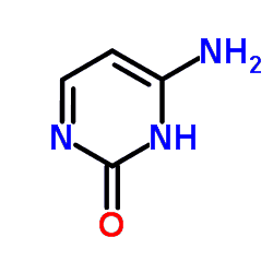
Cytosine
Cytosine
Thymine
Thymine
Uracil
Uracil
    These molecules are nearly flat and relatively insoluble in water. They are joined to sugars at N in pyrimidine bases and a N9 in purine bases by beta-N glycosidic linkage.

Modified Bases

A few modified pyrimidine and purine bases occur in some nucleic acids. They are the post-replicational modifications in DNA and post-transcriptional modification in RNA.
2-Methyladenine
2-Methyladenine
6-Methylaminopurine
6-Methylaminopurine
Nature has created such molecules for its own needs. Some of the viruses make such modifications in order to escape the attack of restriction endonucleases (enzymes). Such modification in t-RNA increases its life and safeguard it from ubiquitously present nucleases.

Sugars

The pentose sugar in DNA in 2’deoxyribose and exists in beta furanose form, whereas in RNA it is ribose (beta-d-ribofuranose). C2’ is endo puckered (out of plane) in DNA, whereas C-3’ endo puckered in RNA. The bases are bonded to C-1’ of 2’-deoxyribose (in DNA and RNA respectively) by beta-N-glycosdic bond. The structural unit containing a base and a sugar is called as nucleoside. The base attachment is of cis type and in anti conformation as shown.
β-D-Ribofuranose
β-D-Ribofuranose
In Nature, the cis and antitype of nucleosides are found only with very rare of trans and syn exceptions. A prime number denotes an atom of the (example 1’) sugar and unprimed number denotes the atoms of the base. The prefix ‘d’ indicates deoxy sugar.

Nucleoside

It is N of pyrimidine base and N9 purine bases joined by beta-N-glycosidic linkage to pentose sugar at C-1’ forming ribonucleoside or deoxyribonucleoside depending upon the type of sugar attached.
Adenosine
Adenosine
Guanosine
Guanosine
Uridine
Uridine
Cytidine
Cytidine
                     

Nucleotide

A molecule containing a base, a sugar, and a phosphoric acid is called a nucleotide. Levenge while working with nucleic acids observed that a phosphate can be attached to 5’ or 3’ position in deoxyribonucleotide and at 5’, 3’ and 2’ position in ribonucleotide. This is because that these positions are vacant or attack positions in nucleosides. In other words, a phosphorylated nucleotide is a nucleotide. If a nucleotide has two or three phosphate groups they are termed as ADP, ATP, GDP, GTP or dATP, dGDP and so on depending upon the type of pentose sugar and number of phosphate groups they have. The ATP is the most important molecule in living organisms as its hydrolysis yields good amount of energy for energy requiring processes. The molecule attains energy during the catabolism and provides energy for anabolism. Later cyclic nucleotides were discovered, which are having great importance in metabolic processes. For example, 3’-5’ cyclic AMP, which is also called as secondary messenger in the cell. Epinephrine and glucagon affect the glycogen metabolism not by entering the cell but by binding at cell plasma membrane and triggering cAMP cascade of reactions.

Functions of Nucleotides

  • Role in energy metabolism, example ATP and GTP molecules
  • Monomer units of ribonuleic and deoxyribonucleic acids.
  • Act as secondary messenger, exammple cAMP.
  • Component of coenzymes viz. NAD, FAD coenzyme A
  • Activated precursor of many physiological reactions i.e. UDP-glucose involved in glycogen synthesis.
  • Allosteric effector of many regulatory reaction in metabolism.

How Nucleotides are Joined to one another.

Levene also made the critical discovery that two nucleotides are linked by covalent phosphodiester bond. Specifically, the 5’-hydroxyl group one nucleotide unit is joined to 3’ hydroxyl group of the next nucleotide by a phosphodiester linkage. Thus the covalent backbone of nucleic acid consists of alternating phosphate and pentose residues. The bases may be regarded as side groups. If two nucleotides are joined by phosphodiester linkage, they are termed as dinucleotide, whereas, when more number of nucleotides are joined, are termed as polynucleotides. Backbone of RNA/ DNA is hydrophilic. The -OH groups of ribose are Hydrogen bonded, whereas the phosphate groups are completely ionized. These negative charges are neutralized with basic proteins, metal ions and polyamines. In eukaryotes they are neutralized by basic proteins called histones. The variable part of DNA is its sequence of four kinds of bases (A, T, G and C) and in RNA (A, C, G and U). The backbone is invariant thoughout the molecule. The structure of DNA chain can be concisely represented in the following way. The symbol for the four principle are deoxyribose nucleosides are: The bold lines refer to the sugar, whereas A, C, G, T denote the bases. The phosphate within the diagonal line the diagram shown below denotes the phosphodiester linkage. The diagonal line joins the end of one bold line and the middle of another. These junctions refer to 5’-oh and 3’ -OH respectively. The 3’ -OH of deoxyadenylate is joined with phosphoryl group at 5’-OH group of deoxycytidine. The basic backbone remains the same throughout, the bases only change. Hence the polynucleotide can also be written as. A DNA chain has polarity. One end of chain has 5’ -OH group and other 3’ -OH group i.e. not linked to another nucleotide. By convention ACGT mean that the unlinked 5’-OH group is on deoxyadenosine, whereas unlinked 3’-OH group on deoxythymidine. The base sequence is written in 5’—> 3’ dirction. As polypeptide. This peptide is not the same as cyc gly ala. Similarly, the base sequence written as ACGT is not the same as TGCA.  

What are essential nutrients for plants

0
Introduction Mineral Metabolism may be defined as a process in which mineral nutrient elements are incorporated into animal/plant metabolites. It’s a known fact that these mineral are got from the soil plants through their roots. Only during the first half of the nineteenth century, plant scientists began to understand the plant growth, development, and need of the chemical elements. These elements are absorbed from soil as inorganic ions. Inorganic ions in the soil are derived mostly from mineral constituents. Due to this reason, we term it as mineral nutrition. Essential Elements Sixteen elements are known to be essential for higher form of life in terms of relative numbers with respect to molybdenum. The level of essential elements known to be critical for the growth of multicellular plants. All the elements present at concentration of 1000ppm or higher are termed macro nutrients (k, Ca, Mg, N, P, S, C, H, O). Potassium, Calcium, and Magnesium are present in soil as cations (K+, Ca++, Mg++) whereas nitrogen, phosphorous and sulfur are normally present in soil as anions (NO3, H2PO4-, SO42-) except C, H, O all the other essential elements are mineral elements. The micronutrients as the name suggests, are required in very small amounts but are equally essential for plant growth (Mo, Cu, Zn, Mn, Fe, B, Cl). In addition to these elements, other elements have been established as being essential for few species of higher plants e.g. Co, Si, Se are important for some higher plants. Specific Functions of Essential Nutrients Since each essential element performs one or more specific metabolic roles, a less than adequate supply of an essential element will be accompanied by distinctive metabolic disruptions, including change in activity of enzymes, in rate of metabolic reactions and concentration of metabolites. This also leads to characteristic changes in the physical appearance of plants, leaves, stem, fruits etc. 1. Sulfur: The higher plants take sulfur as sulfate ion by roots or may be absorbed as SO2 from atmosphere and it is assimilated by leaves. It is a component of cystine, cysteine, methionine and thus proteins, lipoic acid and coenzyme A, TPP, Glutathione, and biotin. 2. Phosphorous: Absorbed (HPO4- or H2PO4-) ion from the soil, the assimilation of phosphorus into ATP is through oxidative phosphorylation Later this inorganic phosphate becomes a part of many molecules of the cell. For example, all the intermediates of glycolysis are phosphorylated. It is also an important component of nucleic acids (as phosphodiester linkage). 3. Magnesium: It serves as a structural component of cell and is involved as a cofactor in many enzymes systems. It is required by the photosynthetic cell containing chlorophyll. It is associated with phosphate transferring reaction. Mg++ forms stable complexes with ATP, ADP, and AMP. 4. Calcium: Calcium has been associated with the cell wall structure as calcium pectate. It is also involved in IAA (Indole acetic acid) stimulated cell elongation and cell division. Nodulation and successful symbiotic nitrogen fixation require relatively high concentration of calcium. Important in enzymic systems viz. Barley alpha-amylase, ATPase and phospholipase-D. 5. Potassium: Many enzymes require potassium for maximum activity viz. aldose (glycolysis), Pyruvate kinase, succinyl CoA synthetase, etc. Translocation of carbohydrates, stomatal opening, and osmotic regulation are affected by its deficiency. 6. Molybdenum: Molybdenum has been shown to be essential for fixation of nitrogen by azotobacter chroccum. It is a constituent of fungi, bacteria and higher plants and it is also a constituent of xanthine oxidase and aldehyde oxidase. 7. Copper: It is found in enzymes in which oxygen is used directly in the oxidation of substrate, e.g., tyrosinase, laccase, ascorbic acid oxidase. It is also involved in oxidation of cytochrome oxidase. 8. Zinc: It is involved in many enzymes, viz, alcohol dehydrogenase, glucose 6-phosphate dehydrogenase, and triose phosphate dehydrogenase. It is highly involved with DNA and helps in regulation of its expression through zinc finger. 9. Manganese: It can replace magnesium in many of the reactions if found insufficient, eg. glucokinase, hexokinase etc. Many citric acid cycle enzymes require manganese for optimal activity. Iron: It is a structural component of porphyrin ring and acts as the cofactor of many enzymatic reactions. In animals, it is associated with porphyrin ring of haemoglobin. It is also a component of electron transport chain, cytochrome b, b6, c, C2, and f. It is also a component of nonheme protein molecule, ferredoxin and in enzyme viz. cytochrome oxidase.

Landforms made by glaciers

0

Glacial Landforms or Landforms made by glaciers

Davis Suggested that a glacial topography is a climatic accident that happens to normal cycle of erosion, i.e., climate gets very cold and the river freezes. Instead of rivers of water there are rivers of ice, called glaciers, operating as the main geomorphic agent. The geological action of glaciers, i.e., erosion, trasportation, and deposition together constitute, what is knows as glaciation.

Formation of Glaciers:-

Under the influence of pressure and moisture, the snowflakes change into a granular ice mass, known as neve in French and ‘firn’ in German. When the ice becomes so thick that the lower layer becomes plastic, outward or downhill flow commences and an active glacier comes into being.

Types of Glaciers. There are three major types of glaciers:

i) The valley glaciers (Mountain glaciers or Alpine glaciers).

Piedmont glacier- these are intermediate in form and origin, between valley glacier and ice-sheet. They are formed at the foot of the mountains.

Ice Sheet.

These are huge covers of ice and are also known as Continental glacier. The largest and thickest glacier are the ice sheets. The Antarctica and Greenland represent more than 95 % of the world’s glacierized surface. Ice sheets are character- ised by a dome-like topography and a general outward flow, barely controlled by the buried subglacial landscape due to the high thickness of the ice. The maximum thickness of ice in Antarctica and Greenland is around 4.7 km and 3 km, respectively. Accordingly, these are two types of glacial topography, one related to the valley glaciation and the other to the continental glaciation.

The topography of valley glaciation

There are two sets of features resulting from glacial erosion and glacial deposition, (since the rock wastes and other loads are carried frozen within the ice during their transportation, in the case of glaciers).

Erosion:

Plucking- Erosion by glaciers take place due to i) plucking ii) rasping and iii) avalanching. Also Read What is geomorphology? Geography Notes

Plucking

Plucking is also known as frost wedging or quarrying. Glacial deepening is mainly accomplished by plucking. During the summer months, the surface parts of a glacier may partially melt. This melts water or rain water down along the sides of the ice mass, finding its way into the cracks and fractures in the rocks along the edges and at the head of the glacier. At night or when the temperature drops this water freezes. It breaks up the rock by frost action, and with the movement of the glacier, they are frozen in suspension in the ice.

Rasping

Rasping is the term used to describe the scraping or abrasion by glacial action. The frost edge of glacier function as bulldozer, pushing and scraping the ground in front of the ice.

Avalanching

Avalanching is the process of mass-wasting. Along the margins of a valley-glacier the valley sides are scrapped and blocks are broken off which become frozen of the sides of the valley and pave the ground for slumping, sliding and debris avalanching which bring great quantities of rock-waste on to the top surface of the glacier.

Feature of Erosion

1. Cirques: amphitheater- These are circular depression formed by plucking and grinding on the upper parts of the mountain-slopes. These are also known as Amphitheater’. 2. Arete. This name is applied to the sharp ridges produced by glacial erosion. Where two cirque-walls intersect from opposite sides, a jagged, knife-like edge, called an ‘arete’ results. It is also known as ‘comb’ or serrate-ridge’. 3. Horn: – Where three or more cirques grow together, a sharp-pointed peak is formed by the intersection of the aretes. Such peaks are known as ‘horns’. 4. Col. Where opposed cirques have intersected deeply, a pass or notch, called a ‘col’ is formed. 5. Glacial-trough. Glacier flow constantly deepens and widens its channel so that after the ice has finally disppeared there remains a deep, steep walled, ‘U’-shaped valley, known as glacial trough. 6. Hanging valley:- Tributary glacers also curve ‘U’-shaped troughs. But they are smaller in cross-section with floors lying high above the floor level of the main-trough, i.e., main glacial valley. Such valleys are called hanging-valleys. 7. Fiords. When the floor of a glacial trough open to the sea lies below sea-level, the sea-water will enter as teh ice front recedes, producing a narrow estuary, known as a ‘fiord’ or fiords’. 8. Tarns. The bed rock is not always evenly excavated under a glacier, so that floors of troughs and cirques may contians rock basin and rock steps. Cirques and upper parts of troughs that are occupied by small lakes, called tarns.

Depositional features of valley-glaciers

Deposition by a glacier takes place when the ice begins to melt and the glacier slow down and vanishes, losing its transporting power. The unstratified, unsorted debris dropped more or less in a random fashion by glaciers form deposits known as morains. Three types of moraines are known, lateral, medial or median, and terminal or end. These three types are differentiated on the basis of their location in the valley. a) Lateral moraine. Deposits of ridge like pattern formed along teh margins of the glaciated valley are known as lateral moraines. b) Medial moraine: – It results due to coalescence of two lateral moraines, where two ice stream join. c) Terminal moraine: – These are accumulation of rock debris at the terminus of a glacier. d) Recessional moraine: Where glacier retreats in a halting manner, a series of concentric moraines is formed, known as ‘recessional moraines’. The topography of ‘continental glaciation’. Like valley glaciers, the continental glaciers proved to be highly effective eroding agent. But continental glacier erodes only by plucking and rasping methods, but erosion process, like avalanching, is absent in case of continental glaciers.

Features of Erosion.

i) Striations: The slowly moving ice scraped and grouped away much solid bed-rock. Left behind were smoothly rounded rock masses bearing countless minute abrasion marks, scratches, called striations. ii) Roches mountonnees: they consist of asymmetrical mounds of rock of varying size, with a gradual smooth abraded slope on one side and a steeper rougher slope on the other. The ‘stoss side’, i.e. the side from which the ice was approaching is characteristically smoothly rounded and the other side, i.e., the ‘lee side’ where the ice plucked out angular joint blocks, is irregular and blocky. They are also known as ‘sheep-rocks’. iii) Crag and tail. Sometimes very hard rocks like volcanic plugs offer great resistance to the ice-flow and stand as pillars in the glaciated valley. These structures are called crags and the lee side which is sloping, in this case, is the tail. Depositional features: The term glacial drift includes all varieties of rock debris deposited in close association with glaciers. These deposits may be classified into two groups 1) Stratified drift, consists of layers of sorted and stratified clay, silts, sands etc. deposited by the meltwater streams and are also known as Glacio-fluvial deposits. 2. Till. It is a heterogeneous mixture of rock fragments ranging in size from clay to boulder which are unsorted and unstratified. A consolidated till is called ‘tillite’. The various depositional features are as follows: a) Drumlin. It consists of glacial till, which is a low mound of clay containing cores of bedrocks. Uphill sides are blunt and down-hill sides are smooth and gently sloping. The long axis of each drumlin parallels the direction of ice-movement and thus serve as indicator sof direction of ice movement. b) Basket of egg-topography:- The drumlins commonly occur in groups of swarms, which may number in the hundreds; the topography produced by them is peculiar and is known as basket of egg topography. c) Ground moraine: – Between moraines, the surface overridden by the ice is overspread by a cover of glacial till, known as ground moraine. Thus it is the sheet of debris left after a steady retreat of ice. Flacio-Fluvial Deposits: i) Outwash plain:- It is also known as overwash plain. Glacial streams carry a huge quantity of rock-debris and then form fan-like plains beyond the terminus of glaciers. These are stratified. When they occur on valley floors such outwash plains are called ‘valley trains’. ii) Kames or kame terraces: These are formed on the top surface of the glacier where the surficial melt-water wash sediments from the top into depressions. As the ice melts the material that formerly filled depressions on top of the glacier is dropped and makes small hills, which are more or less flat-topped and are known as kames. Terraces, called kame terraces, are built in this way. iii) Eskers: These are winding steep-sided ridge like features built of stream borne drift. These are known as Osser or Oss. Erratics: These are stray boulders of rocks which have undergone a prolonged glacial transport and have subsequently been deposited in an area, where the country rocks are of distinctly different types. At times they are delicately balanced upon glaciated bed rock, and are called poking or logging-stone. Kettles: – Drifts occurring in the vicinity of a glacier and particularly those lying near about the ice-terminus are ordinarily found to contain a number of depressions, some of which may give rise to lakes or swamps. Such hollows are known as kettles vi) Varves:- These are layered clays alternating with coarse and finer sediments. Other Important Features associated with Glacier: i) Nivation: from – It is the process of quarrying of rocks mostly by frost action ii) Ablation: – It includes process both evaporation and the melting of snow and ice iii) Calving: – Within fiords, glaciers come in contact with marine water and blocks of ice are found to break from the mass of the glacier. This process of wastage of glacier is known as calving. Serace: – Similar to a waterfall in a river, in a steeper section of the valley, the glacier is broken up into rugged ice-pinnacles and is known as serace. Iceberg: – These are floating ice-hill on the sea-water. Crevasses: – These are cracks formed due to differential movement within the mass of the glacier. In German, they are known as bergschrund Nunatak: nunatak- A rock mass which projects through an ice-sheet, generally found at the margins of a sheet where the ice is thinnest, is nunatak.

Cell cycle and their different phases | Questions | Biology Notes

0

Cell Cycle and Cell Division Biology Notes

Introduction of Cell Cycle

Cell Cycle : All organisms start their life from a single cell and grow by the addition of new cells. The new cells arise by the division of pre-existing cells. This idea was suggested by Rudolf Virchow in 1858 in a particular statement ‘Omnis Cellula e Cellula’, means every cell produces from a cell. This states that the continuity of life depends on cell reproduction or cell division.

Who introduced the Cell cycle?

Cell cycle was introduced by Howard and Pele in 1953. It is defined as the series of events by which a cell duplicates its genome and synthesizes other cell components and then divides into two daughter cells

Phases of cell cycle

Cell cycle occurs in the following two phases.
  1. Interphase (nondividing phase)
  2. M Phase or Mitosis Phase (dividing phase)

Interphase

It represents the phase between two successive M phases. It constitutes or lasts for more than 95% of the whole duration of the cell cycle. Though it is called the resting phase, it is the time during which the newly formed cells prepare themselves for division i.e., to undergo both growth and DNA replication in an orderly manner.

M Phase

It is the phase of cell division in which already duplicated chromosomes get distributed into two daughter nuclei. It starts with the nuclear division (karyokinesis) and terminates after cytokinesis Karyokinesis is the separation of daughter chromosomes and nucleus division and cytokinesis is the division of cytoplasm. During this phase, all components of the cell reorganise for cell division. Since, the number of chromosomes remains same in both parent and progeny cells, it is also known as equational division.

Cell Division

It is a very important phenomenon in all living organisms. The concept of cell division was firstly propounded by a scientist Nageli and was observed by Flemming in 1882 in reptelean Triturus mascules and gave it a name mitosis. Its complete extensive and exclusive study and was done by Belar in 1920. This is also called cell production.

Modes of Cell divison

Cell division usually occurs in following three ways
  1. Amitosis
  2. Mitosis
  3. Meiosis
Amitosis
It is very rare and is not considered an exact mode of cell division. It occurs only in some specialised cells like mammalian cartilage, embryonic membrane of some vertebrates, old tissues, diseased tissues, etc.
Mitosis
It was first explained by Eduard Strasburger. It usually takes place in somatic cells of animals Thus, it is known as somatic division. Mitosis occurs in gonads for the multiplication of undifferentiated germ cells. It is continuous process that gives rise to two identical cells but the number of chromosomes in them remains the same. It occurs in various phases such as prophase, metaphase, anaphase, telophase and then cytokinesis. Cell cycle cell division mitosis meiosis

The significance of Mitosis

It helps in the growth and development of multicellular organisms, in the healing and repair of wounds; in maintaining the chromosome number and nucleocytoplasmic ratio, etc.

Meiosis

The term meiosis was given by JB Farmer and Moore in 1905. Meiosis as division process is restricted to only reproductive cells due to which gametes (sex cells) are produced. It occurs at a particular time during which a diploid cell divides to give rise to four haploids cells. It basically produces gametes in animals, some lower plants, various protists, and fungi. Meiosis is asexually reproducing organisms form asexual reproductive bodies like spores. As meiosis results in the reduction of the number of chromosome in the daughter cells by half, so it is also known as reduction division. It consists of two stages of division that occur successively in an organism with one time chromosome replication. First Meiotic Division (Meiosis I) Second Meiotic Division (Meiosis II)

Meiosis I

In this phase of division, parental chromosomes replicate to produce identical sister chromatids and the number of chromosomes reduces from diploid (2n) to haploid (n) and hence, this type of division is called heterotypic division. Like mitosis, it also involves the four phases of division as described below. Prophase I:- The prophase I of meiosis is more longer thatn the prophase of mitosis and it takes more than 90% of time required for meiosis. Prophase I is further divided into 5 sub-stages such as leptotene, zygotene, pachytene, diplotene and diakinesis Metaphase I during the course of this phase, spindle shifts to the position formerly taken by the nucleus and the synapsed pair of chromosome (bivalent) get arranged around the equator of the spindle and are attacked by their centromere. Anaphase I:- During anaphase, I, homologous chromosomes of each pair gets separated and half of the chromosome move to each pole. Reduction of chromosome occur and each chromosome at individual poles is still double and have two chromatids Telophase I The arrival of homologus chromosome at opposite pole shows the end of meiosis I. During this phase, chromosomes uncoil and get elongated. Cytoplasm tends to get divided by cleavage (constriction) in an animal cell and by cell plate formation in a plant cell and produces two cells each with one nucleus. Meiosis II The meiotic division exactly the same in overall process as mitotic division. There is no reduction in the number of chromosomes and the haploid nuclei divide mitotically in order to produce four haploid daughter nuclei. Thus, each diploid nucleus which undergoes meiosis produces four haploid nuclei. The only difference between mitosis and meiosis II is that interphase do not proceed prophase in meiosis. It gets initiated immediately after cytokinesis, usually before the chromosomes have been fully elongated After meiosis II, four daughter cells are formed from the original single parent cell and each one is haploid (n) in nature. The phases involved in meiosis II are prophase Ii, metaphase II, anaphase II and telophase II. Significance of Meiosis: – Meiosis is significantly proved to be the important mechanism in living organism because this process brings stability in the number of chromosome in an organism. It also increases genetic variability in the population of organism from one generation to next. As variations are important to the process of evolution, meiosis acts as a source of new genetic variation.

Questions related to Cell cycle and cell division which will helps to more deeply clear the topics

Where in the cell cycle does apoptosis occur?

In the normal pathway, DNA damage is an intracellular signal that is passed via 2 protein kinases, leading to activation of p53. Activated p53 promotes 4 transcription of the gene for 5 a protein that inhibits the cell cycle. The resulting suppression of cell divisionensures that the damaged DNA is not replicated. If the DNA damage is irreparable, then the p53 signal leads to programmed cell death called apoptosis.

What controls the cell cycle?

The cell cycle control system has been compared to the control device of a washing machine. Like the washer’s timing device, the cell cycle control system proceeds on its own, according to a built-in clock. However, just as a washer’s cycle is subject to both internal control (such as the sensor that detects when the tub is filled with water) and external adjustment (such as starting or stopping the machine), the cell cycle is regulated at certain checkpoints by both internal and external signals. A checkpoint in the cell cycle is a control point where stop and go-ahead signals can regulate the cycle. Three important checkpoints are found in the G 1 , G 2 , and M phases.

Which cell cycle phase takes the longest?

G1 is typically the longest phase of the cell cycle.

Which cell cycle phase is the shortest?

Mitosis

When during cell cycle are chromosomes visible?

Mostly chromosomes are not visible. The chromosomes are visible during the mitosis in the cell cycle

Why is Cell Division Important?

Cell division is necessary for the growth of organisms, for wound healing, and to replace cells that are lost regularly, such as those in your skin and in the lining of your gut.

What cell division is asexual?

The small organism like bacteria reproduced by the asexual method of reproduction. Thus all the growth in bacterial population is clonal, it’s division of existing cells to new one. No fertilization take place in this type of reproduction. The new daughter cell which is produced having the same genetic information as of their parents.

What cell division process is responsible for growth?

Mitosis cell division helps in the growth and development of multicellular organisms; in the healing and repair of wounds; in maintaining the chromosome number and nucleocytoplasmic ratio, etc.

How is cell cycle regulated?

The frequency of cell division varies with the type of cell. For example, human skin cells divide frequently throughout life, whereas liver cells maintain the ability to divide but keep it in reserve until an appropriate need arises say, to repair a wound. Some of the most specialized cells, such as fully formed nerve cells and muscle cells, do not divide at all in a mature human. These cell cycle differences result from regulation at the molecular level. The mechanisms of this regulation are of great interest, not only to understand the life cycles of normal cells but also to learn how cancer cells manage to escape the usual controls

How are mitosis and binary fission different?

Binary fission is taking place mainly in the prokaryotes and single cell organism like bacteria. Mitosis mainly occurs in multicellular Eukaryotes. The term binary fission, meaning “division in half,” refers to this process and to the asexual reproduction of single-celled eukaryotes, such as the amoeba. Mitosis consists of multiple phases: prophase, prometaphase, metaphase, anaphase, and telophase. Binary fission is not complex like the Mitosis.

Where is Mitosis in the cell cycle?

The cell cycle is divided into the five phases named, G1 (gap phase), S (synthesis), G2 (gap phase 2), Mitosis, Cytokinesis. as shown in the Fig.

Which cells undergo mitosis?

Mitosis occurs in almost every cell of eukaryotes. It occurs where the need of the number of cells is more. Whereas meiosis is occurred only in special cells, in animals, gametes, and in plants pollen.

Who discovered mitosis in animals?

When Flemming looked at the cells through what would now be a rather primitive light microscope, he saw minute threads within their nuclei that appeared to be dividing lengthwise. Flemming called their division mitosis, based on the Greek word mitos, meaning “thread.” But Otto Bütschli might have claimed the discovery of the process presently known as “mitosis”

What mitosis stage is the longest?

Prophase is the longest phase of mitosis, but it occurs faster than interphase

What mitosis and meiosis have in common? or Similarities between mitosis and meiosis

S.No.

Characters

Mitosis

Meiosis
I. General
(1) Site of occurrence Somatic cells and during the multiplicative phase of gametogenesis in germ cells. Reproductive germ cells of gonads.
(2) Period of occurrence Throughout life. During sexual reproduction.
(3) Nature of cells Haploid or diploid. Always diploid.
(4) Number of divisions Parental cell divides once. Parent cell divides twice.
(5) Number of daughter cells Two. Four.
(6) Nature of daughter cells Genetically similar to parental cell. Amount of DNA and chromosome number is same as in parental cell. Genetically different from parental cell. Amount of DNA and chromosome number is half to that of parent cell.
II. Prophase
(7) Duration Shorter (of a few hours) and simple. Prophase-I is very long (may be in days or months or years) and complex.
(8) Subphases Formed of 3 subphases : early-prophase, mid-prophase and late-prophase. Prophase-I is formed of 5 subphases: leptotene, zygotene, pachytene, diplotene and diakinesis.
(9) Bouquet stage Absent. Present in leptotene stage.
(10) Synapsis Absent. Pairing of homologous chromosomes in zygotene stage.
(11) Chiasma formation and crossing over. Absent. Occurs during pachytene stage of prophase-I.
(12) Disappearance of nucleolus and nuclear membrane Comparatively in earlier part. Comparatively in later part of prophase-I.
(13) Nature of coiling Plectonemic. Paranemic.
III. Metaphase
(14) Metaphase plates Only one equatorial plate Two plates in metaphase-I but one plate in metaphase-II.
(15) Position of centromeres Lie at the equator. Arms are generally directed towards the poles. Lie equidistant from equator and towards poles in metaphase-I while lie at the equator in metaphase-II.
(16) Number of chromosomal fibres Two chromosomal fibre join at centromere. Single in metaphase-I while two in metaphase-II.
IV. Anaphase
(17) Nature of separating chromosomes Daughter chromosomes (chromatids with independent centromeres) separate. Homologous chromosomes separete in anaphase-I while chromatids separate in anaphase in anaphase-II.
(18) Splitting of centromeres and development of inter-zonal fibres Occurs in anaphase. No splitting of centromeres. Inter-zonal fibres are developed in metaphase-I.

V. Telophase

(19) Occurrence Always occurs Telophase-I may be absent but telophase-II is always present.
VI. Cytokinesis
(20) Occurrence Always occurs Cytokinesis-I may be absent but cytokinesis-II is always present.
(21) Nature of daughter cells 2N amount of DNA than 4N amount of DNA in parental cell. 1 N amount of DNA than 4 N amount of DNA in parental cell.
(22) Fate of daughter cells Divide again after interphase. Do not divide and act as gametes.
VII. Significance
(23) Functions Helps in growth, healing, repair and multiplication of somatic cells. Occurs in both asexually and sexually reproducing organisms. Produces gametes which help in sexual reproduction.
(24) Variations Variations are not produced as it keeps quality and quantity of genes same. Produces variations due to crossing over and chance arrangement of bivalents at metaphase-I.
(25) In evolution No role in evolution. It plays an important role in speciation and evolution.
 

How mitosis relates to cancer?

Without mitosis, their is no cancer. In cancer cells grows or divided at abnormal speed and resulted in tumor formation. The cells are divided by the process named ‘mitosis’.

When mitosis occurs without cytokinesis?

Cytokinesis is the last phase of the mitosis, in which cell is divided into two parts. In mitosis, there is replication of chromosome and two number of the nucleus are formed for two different cell. If, the cytokinesis does not take place then the cell results with two nuclei. Such a cell is called a multinucleated cell. This can be a normal process. For example, humans have certain multinucleated bone cells (osteoclasts) that are formed this way. Mitosis without cytokinesis is also observed in the early development of certain insects such as the fruit fly (Drosophila).

Why is mitosis known as equational division?

Mitosis is the process of cell division gives 2 number of daughter cells. The chromosome number in each daughter cell is equal to that in the parent cell, i.e., diploid. Hence, mitosis is known as equational division.

New research could help humans see what nature hides

0

Things are not always as they appear. New visual perception research at The University of Texas at Austin, published in the Proceedings of the National Academy of Sciences, explains the natural limits of what humans can see and how to find what nature hides.

UT Austin researchers investigated the three main background properties that affect the ability to see objects: the luminance or brightness, the contrast (the variation in luminance) and the similarity of the background to the orientation and shape of the object. Using an experimental and theoretical approach involving analysis of millions of natural images, the researchers found that the ability to detect the differences between the object and the background was predicted directly from the physics of natural stimuli. “The discovery of highly systematic laws for perception in natural scenes — made possible by constrained statistical sampling — is a potential game changer,” said the paper’s lead author Wilson Geisler, a UT Austin professor of psychology and director of the Center for Perceptual Systems. “It demonstrates how to study complex real-world perception with the same level of rigor that was previously achieved only with simple synthetic stimuli.” In order for people to pick out an object against a background, the object must differ from the background by a “just noticeable difference,” a threshold quantified by the minimum difference a person can detect the majority of the time. Even as the properties of both the object and the background vary, the threshold remains in constant proportion to the product of the background properties — a generalized version of Weber’s law. “The ability of these background properties to mask objects is well known for simple laboratory stimuli,” Geisler said. “However, it was not known how these properties combine to mask objects in natural scenes.” The researchers considered the effects of stimulus uncertainty. Under real-world conditions, the properties of the object and of the background against which the object appears will randomly vary from one occasion to the next, creating a stimulus uncertainty that can also affect accuracy in detecting the object. Their findings showed that the detrimental effects of this uncertainty can be minimized by estimating the luminance, contrast and similarity at the object’s possible locations, and then dividing the neural responses at each of these locations by the product of these estimates. The researchers found strong evidence that these computations are done automatically in the human visual system. Knowing this may lead to improved radiology technology to help radiologists identify abnormalities in the human body; or better security imaging at airports to detect suspicious items in luggage; or enhanced camouflage design to disguise soldiers in war zones, Geisler said. “There are many potential applications of these findings. For example, radiological images are highly complex, like the natural images that drove the evolution of the human visual system. Thus, the perceptual laws for natural images may predict when a radiologist will have difficulty detecting suspicious objects in a radiological image. These predictions could be used to alert the radiologist to locations where extra scrutiny would be advised,” Geisler said.

Don’t leave baby boomers behind when designing wearable technology

0

Wearable devices have been heralded as one of the next great technological frontiers. They can provide all users, including older ones, with constantly updated medical information by tracking cardiac health, identifying potential illnesses, and serving as emergency alert systems, among other benefits.

That is, if you can get older users to adopt the wearable technology. In their article in the July 2017 issue of Ergonomics in Design, “Designing Wearable Technology for an Aging Population,” human factors/ergonomics researchers lay out a framework for improving the usability of wearable technology for older adults. According to Joanna Lewis, a doctoral student of applied experimental and human factors psychology at the University of Central Florida, “The proportion of the population over the age of 65 is growing and will continue to do so. Technological developments are exponentially growing and inundating our lives, and we don’t want a demographic that is scaling up in size not to have access to devices that are becoming prolific in everyday society.” Although wearable devices can serve as important tools for older adults, Lewis and coauthor Mark Neider found that poor design decisions that fail to address the aging population’s needs can undermine the technology’s value. Older adults also tend to experience feelings of mistrust and frustration when using new devices, with the result that they often abandon otherwise worthwhile technology. Taking into account the role of age-linked declines in cognitive, physical, and sensory abilities, the authors identified several critical areas for improvement. These include reducing the steps required for users to complete a given action, minimizing the need for multitasking, eliminating time constraints for completing a task, and increasing the size of buttons, icons, and text. Lewis and Neider also caution designers to avoid clunky or outdated exteriors that may result in age-related stereotypes or cause users to feel stigmatized by their peers. “A device’s usability should consider all ages,” Lewis adds. “Potential issues with wearable devices for older adults can be avoided by acknowledging limitations, and development teams can create effective and safe platforms that appeal to a variety of end users.”

Could this strategy bring high-speed communications to the deep sea?

0

A new strategy for sending acoustic waves through water could potentially open up the world of high-speed communications activities underwater, including scuba diving, remote ocean monitoring, and deep-sea exploration.

By taking advantage of the dynamic rotation generated as acoustic waves travel, the orbital angular momenta, researchers at the Department of Energy’s Lawrence Berkeley National Laboratory (Berkeley Lab) were able to pack more channels onto a single frequency, effectively increasing the amount of information capable of being transmitted. They demonstrated this by encoding in binary form the letters that make up the word “Berkeley,” and transmitting the information along an acoustic signal that would normally carry less data. They describe their findings in a study published this week in the Proceedings of the National Academy of Sciences. “It’s comparable to going from a single-lane side road to a multi-lane highway,” said study corresponding author Xiang Zhang, senior faculty scientist at Berkeley Lab’s Materials Sciences Division and a professor at UC Berkeley. “This work has a huge potential in high-speed acoustic communications.” While human activity below the surface of the sea increases, the ability to communicate underwater has not kept pace, limited in large part by physics. Microwaves are quickly absorbed in water, so transmissions cannot get far. Optical communication is no better since light gets scattered by underwater microparticles when traveling over long distances. Low frequency acoustics is the option that remains for long-range underwater communication. Applications for sonar abound, including navigation, seafloor mapping, fishing, offshore oil surveying, and vessel detection. However, the tradeoff with acoustic communication, particularly with distances of 200 meters or more, is that the available bandwidth is limited to a frequency range within 20 kilohertz. Frequency that low limits the rate of data transmission to tens of kilobits per second, a speed that harkens back to the days of dialup internet connections and 56-kilobit-per-second modems, the researchers said. “The way we communicate underwater is still quite primitive,” said Zhang. “There’s a huge appetite for a better solution to this.” The researchers adopted the idea of multiplexing, or combining different channels together over a shared signal, or multiplexing, is a technique widely used in telecommunications and computer networks. But multiplexing orbital angular momentum is an approach that had not been applied to acoustics until this study, the researchers said. As sound propagates, the acoustic wavefront forms a helical pattern, or vortex beam. The orbital angular momentum of this wave provides a spatial degree of freedom and independent channels upon which the researchers could encode data. “The rotation occurs at different speeds for channels with different orbital angular momentum, even while the frequency of the wave itself stays the same, making these channels independent of each other,” said study co-lead author Chengzhi Shi, a graduate student in Zhang’s lab. “That is why we could encode different bits of data in the same acoustic beam or pulse. We then used algorithms to decode the information from the different channels because they’re independent of each other.” The experimental setup, located at Berkeley Lab, consisted of a digital control circuit with an array of 64 transducers, together generating helical wavefronts to form different channels. The signals were sent out simultaneously via independent channels of the orbital angular momentum. They used a frequency of 16 kilohertz, which is within the range currently used in sonar. A receiver array with 32 sensors measured the acoustic waves, and algorithms were used to decode the different patterns. “We modulated the amplitude and phase of each transducer to form different patterns and to generate different channels on the orbital angular momentum,” said Shi. “For our experiment we used eight channels, so instead of sending just 1 bit of data, we can send 8 bits simultaneously. In theory, however, the number of channels provided by orbital angular momentum can be much larger.” The researchers noted that while the experiment was done in air, the physics of the acoustic waves is the very similar for water and air at this frequency range. Expanding the capacity of underwater communications could open up new avenues for exploration, the researchers said. This added capacity could eventually make the difference between sending a text only message and transmitting a high-definition feature film from below the ocean’s surface. Remote probes in the oceans could send data without the need to surface. “We know much more about space and our universe than we do about our oceans,” said Shi. “The reason we know so little is because we don’t have the probes to easily study the deep sea. This work could dramatically speed up our research and exploration of the oceans.”

Odd properties of water and ice explained: Water exists as two different liquids

0

We normally consider liquid water as disordered with the molecules rearranging on a short time scale around some average structure. Now, however, scientists at Stockholm University have discovered two phases of the liquid with large differences in structure and density. The results are based on experimental studies using X-rays, which are now published in Proceedings of the National Academy of Science.

Most of us know that water is essential for our existence on planet Earth. It is less well-known that water has many strange or anomalous properties and behaves very differently from all other liquids. Some examples are the melting point, the density, the heat capacity, and all-in-all there are more than 70 properties of water that differ from most liquids. These anomalous properties of water are a prerequisite for life as we know it. “The new remarkable property is that we find that water can exist as two different liquids at low temperatures where ice crystallization is slow,” says Anders Nilsson, professor in Chemical Physics at Stockholm University. The breakthrough in the understanding of water has been possible through a combination of studies using X-rays at Argonne National Laboratory near Chicago, where the two different structures were evidenced and at the large X-ray laboratory DESY in Hamburg where the dynamics could be investigated and demonstrated that the two phases indeed both were liquid phases. Water can thus exist as two different liquids. “It is very exciting to be able to use X-rays to determine the relative positions between the molecules at different times,” says Fivos Perakis, postdoc at Stockholm University with a background in ultrafast optical spectroscopy. “We have in particular been able to follow the transformation of the sample at low temperatures between the two phases and demonstrated that there is diffusion as is typical for liquids.” When we think of ice it is most often as an ordered, crystalline phase that you get out of the ice box, but the most common form of ice in our planetary system is amorphous, that is disordered, and there are two forms of amorphous ice with low and high density. The two forms can interconvert and there have been speculations that they can be related to low- and high-density forms of liquid water. To experimentally investigate this hypothesis has been a great challenge that the Stockholm group has now overcome. “I have studied amorphous ices for a long time with the goal to determine whether they can be considered a glassy state representing a frozen liquid,” says Katrin Amann-Winkel, researcher in Chemical Physics at Stockholm University. “It is a dream come true to follow in such detail how a glassy state of water transforms into a viscous liquid which almost immediately transforms to a different, even more viscous, liquid of much lower density.” “The possibility to make new discoveries in water is totally fascinating and a great inspiration for my further studies,” says Daniel Mariedahl, PhD student in Chemical Physics at Stockholm University. “It is particularly exciting that the new information has been provided by X-rays since the pioneer of X-ray radiation, Wolfgang Röntgen, himself speculated that water can exist in two different forms and that the interplay between them could give rise to its strange properties.” “The new results give very strong support to a picture where water at room temperature can’t decide in which of the two forms it should be, high or low density, which results in local fluctuations between the two,” says Lars G.M. Pettersson, professor in Theoretical Chemical Physics at Stockholm University. “In a nutshell: Water is not a complicated liquid, but two simple liquids with a complicated relationship.” These new results not only create an overall understanding of water at different temperatures and pressures, but also how water is affected by salts and biomolecules important for life. In addition, the increased understanding of water can lead to new insights on how to purify and desalinate water in the future. This will be one of the main challenges to humanity in view of the global climate change.

ICAR JRF (Junior Research Fellowship Exam) | Mock Tests | Study Material

0

ICAR JRF EXAM 

ICAR JRF: Every year ICAR (Indian Council of Agricultural Research) awarded the JRF (Junior Research Fellowship) to Agriculture Students or student having BSc. in Agricultre, Forestry etc.

Amount awarded to Student in ICAR JRF

Rs. 8640/- or 130USD pm + Contingency grant of Rs. 6000/- or 90 USD per year for other than Veterinary Science students Rs. 12000/- or 180 USD pm + Contingency grant of Rs. 6000/- or 90 USD per year for Veterinary Science students Fellowship is given to the number of students for their further Post graduation studies who scored good rank in this exam. So, as we promise to help the every student to fulfill their goal. We will start the providing free study material as well as mock test. We will provide the MOCK Test for Horticulture, Agronomy, Soil Science, Plant Breeding, Plant Protection, Foresty and other subjects. Our mock test covers the entire syllabus of the different subject of the in JRF Exam. Currently, we are not able to provide notes instantly, that’s why we recommend the following books to the students. Give your review about out this initiative by commenting the comment section and become partner in this initiative by contacting us at [email protected]